Your eyes – the window to your soul by Dr Enslin Uys (Ophthalmologist)
There are so many different diseases that can affect the eyes and reduce your vision and quality of life. Many of these diseases are preventable and can be picked up by routine eye examination. I mention only but a few common eye problems: cataract formation; glaucoma; diabetic eye disease; age related macular degeneration (AMD), retinal vein occlusion.
How often should you have your eyes checked? There is no hard or fast rule. If you don’t have any symptoms or vision problems, doctors recommend getting regular eye
exams based on your age:
• Ages 20 to 39: Every 5 years.
• Ages 40 to 54: Every 2 to 4 years.
• Ages 55 to 64: Every 1 to 3 years.
• Ages 65 and older: Every 1 to 2 years
Here are 8 signs that you should get another exam on the calendar soon:
• You are seeing new spots, flashes of light, or floaters.
• You have diabetes or another health condition that affects your eyes. Also, if you have a family history of conditions like diabetes or glaucoma you may need exams more often, especially as you move into your 50’s and beyond.
• You can’t remember when you last had an eye exam. If it’s been longer than a year, you’re overdue.
• You have difficulty driving at night with scattering of light and difficulty seeing street signs in the dark. You have difficulty reading other cars number plates during the day.
• You experience eye strain, headaches and/or blurred vision after spending an extended amount of time in front of a computer screen.
• You get motion sick, dizzy, or have trouble following a moving target.
• You hold books or the newspaper further away from your face and squint or
close one eye to read them clearly.
• You notice any changes in your vision, especially after an incident of head trauma.
Don’t wait until you experience any of these before you schedule an eye exam. Keep in mind that an eye exam benefits more than just your eyes. Your eye doctor can detect a wide range of diseases like diabetes, retinal vein occlusion and glaucoma just by looking at your eyes.
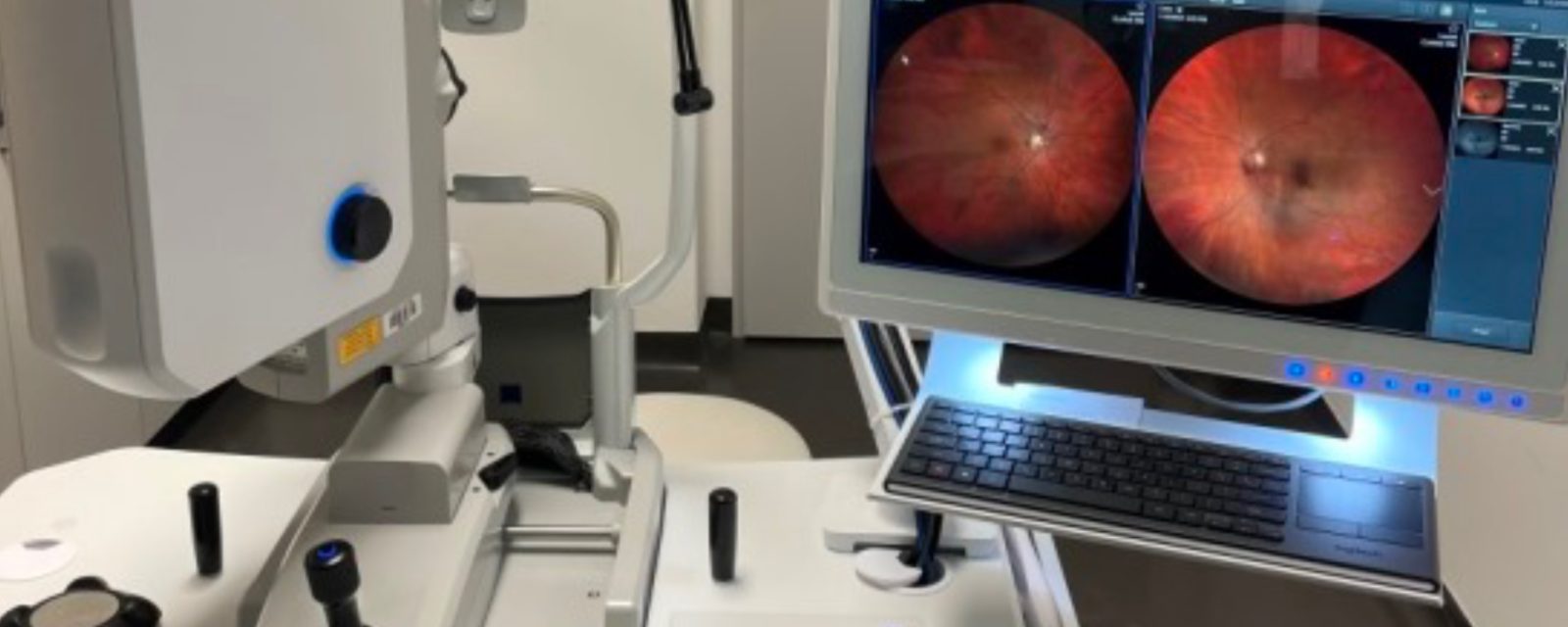
Dr Enslin Uys has recently acquired the state of the art Zeiss Clarus 700 wide angle fundus camera system. Ultra-widefield fluorescein angiography (FA) is a useful exam to identify diabetic retinopathy, especially visualizing the peripheral retina which is fundamental to assess nonperfused areas, vascular leakage, microvascular abnormalities, and neovascularisations. With the ZEISS CLARUS 700 ultra-widefield imaging and high resolution FA, the smallest details from ONH to the periphery can be captured in early phase. Wide field (WF) fundus photography is the most impressive technology Dr Uys have added to his practice since he began using OCT for optic nerve and retinal examinations. The experience from other eye care professionals shows that WF screening photos improve quality of care. Although there were no abnormal findings on the OCT, the peripheral lesions found in an ultra-wide field (UWF) image were concerning as clinical studies have shown that predominantly peripheral lesions are four times more likely to progress to proliferative diabetic retinopathy over four years. Finding these peripheral lesions allows Dr Uys to manage the diabetic retinopathy more effectively and initiate appropriate treatment earlier.


